Minimally Invasive
Surgery Case of the Day
History: A 21 year old
female dancing student presents with back pain and left sided L5 radicular pain
of 3 months durantion.
Findings: Paraspinal
left sided tenderness and a positive left sided straight leg raise test is seen
on examination.
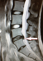
Imaging: MRI reveals a
foraminal left sided L5/S1 herniation (arrows)
Procedure: Transforaminal
L5/S1 endoscopic discectomy is performed
as an out-patient procedure through a 0.5cm incision
Surgeon: George Rappard,
MD
Disposition: The patient is
discharged one hour post-procedure with resolution of radiculopathy and
reversal of straight leg raise sign
Procedure Images:
Blue stained herniated disc material is seen through scope (arrow)
Upon exploring foramen, exiting L5 nerve root (arrow) and
foraminal extruded disc (star) is seen
The endoscopic bipolar (left) and 2.5mm rongeur (right) are
used to remove foraminal herniation.
Arrow denotes exiting and displaced L5 nerve root.
After decompression the annular tear is probed (arrow) for
loose intradiscal fragments
The nerve (arrow) is decompressed and the foraminal herniation
is now seen as an empty cavity (star)
For more information on endoscopic lumber discectomy contact
the Los Angeles Minimally Invasive Spine Institute or go to http://www.lamisinstitute.com/percutaneous-endoscopic-lumbar-discectomy


No comments:
Post a Comment